SingHealth Duke-NUS Academic Medical Centre will NEVER ask you to transfer money over a call. If in doubt, call the 24/7 ScamShield helpline at 1799, or visit the ScamShield website at www.scamshield.gov.sg.
Imaging Capabilities
Imaging Capabilities
For high resolution imaging of lying or awake animals, including 3 types of animal positioning systems as well as imaging of small and medium sized animals.
CT subsystem
- Maximum FOV: ~20 x 15 cm
- Spatial resolution (FWHM): <30µm @ 10% MTF
- Scanning: Circular / helical
- Magnification: X1.5
PET subsystem
- Axial FOV: Single FOV: 15 cm
- Transaxial FOV: 20 cm
- Spatial resolution:
- 1 mm (Tera-Tomo 3D PET reconstruction engine)
- 1.5mm (with FBP according to NEMA standards)
- Temporal resolution: 1.0ns
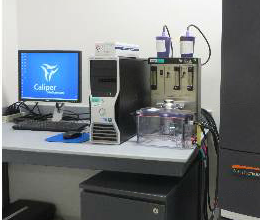
High sensitivity In-vivo optical (fluorescence and bioluminescence) imaging for small animals.
- 3D tomographic reconstruction
- High throughput with 5 mice at 23cm FOV
- High resolution to 20 microns with 3.9cm FOV
- 28 high efficiency filters: 10 narrow band Ex filters (415 - 760nm with 30nm bandwidth)
- Multispectral imaging with superior spectral unmixing properties
- Ideal for distinguishing multiple bioluminescent and fluorescent reporters
- Ability to import and automatically co-register CT or MRI images yielding functional and anatomical context
Real time x-ray imaging of large animals.
- C-arm orbital movement: 132° (-42° to +90°)
- C-arm angulation: ±190°
- C-arm horizontal movement: 20 cm
- C-arm immersion depth: 73 cm
- C-arm swivel range: ±10°
- C-arm vertical movement: 38 cm, motorized
- Camera with CCD sensor: 1034 (H) x 1024 (V)

Portable Real time x-ray imaging of large animals.
- Footprint (L x W x H): 131cm x 63cm x 133cm
- Max. focal spot height: 190cm
- Max. horizontal tube extension: 110cm

For non-invasive image acquisition of large animals.
- Frequency: 50/60 Hz
- Operating modes: B-mode, M-mode, Color flow, Power Doppler Imaging (PDI), PW & CW Doppler, 2D volume mode
- Horizontal/vertical viewing angle of +/- 170 ̊
© 2025 SingHealth Group. All Rights Reserved.


